Partitioning tissues into compartments that do not intermix is essential for the correct morphogenesis of developing animal tissues. However, the mechanisms that keep cells apart at compartmental boundaries remain unclear.
We have shown in Drosophila embryos that actomyosin-based barriers stop cells from invading the adjacent compartment [1]. Actomyosin cytoskeletal components are enriched at compartmental boundaries, forming cable-like structures which are not dismantled in dividing cells. When non-muscle myosin II (Myo II) function is inhibited directly at these cables using CALI, dividing cells close to the boundary invade the wrong compartment. We have proposed that local regulation of actomyosin contractibility is the primary mechanism sorting cells at compartmental boundaries.
Myosin II activity is essential for several common morphogenetic mechanisms in Drosophila and vertebrate tissues, such as apical constriction during tissue internalization or bending, and cell intercalation during body axis extension [2]. Boundary formation is thus another example where intrinsic forces produced by Myosin II activity sculpt developing tissues.
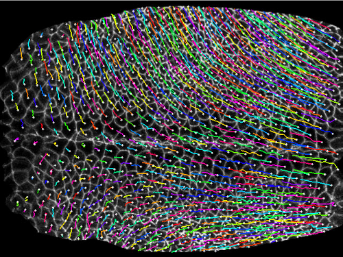
In addition to intrinsic forces, extrinsic forces (produced by deforming tissues or fluid flows) are likely to be important in morphogenesis. We have found evidence that an extrinsic axial force extends the main body axis in Drosophila embryos, acting in parallel to Myo II-dependent active cell intercalation [3,4]. The axial force elongates ectodermal cell shapes along the main body axis, this elongation becoming more severe in absence of active cell intercalation. Axial cell elongations are diminished in absence of mesoderm invagination, suggesting that this morphogenetic movement produces a force that propels axis extension.
References:
[1] Monier et al. Nat Cell Biol (2010) vol. 12 pp. 60-5.
[2] Lye & Sanson, Curr Top Dev Biol (2011) vol. 95 pp. 145-187.
[3] Butler et al. Nat Cell Biol (2009) vol. 11 pp. 859-64.
[4] Blanchard et al. Nat Methods (2009) vol. 6 pp. 458-64.